 |
A Drexel University team of engineers, scientists and biologists have developed a carbon nanotube-based device for probing single living cells without damaging them. ...
Multifunctional carbon-nanotube cellular endoscopes
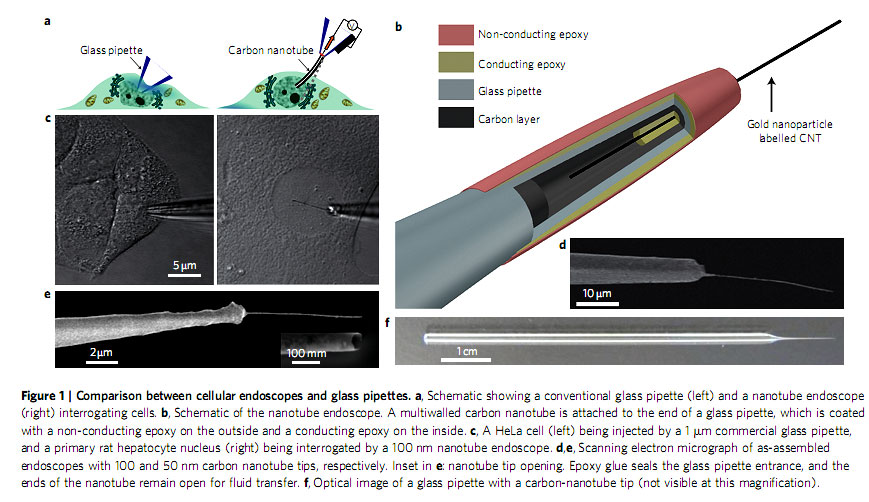
Artist renderings of a nano-needle poking a single cell have become the symbol of nanotechnology, surfacing on covers of magazines and books for about a decade but actual nano-needles able to interrogate small cells without causing cellular damage have not become reality until recently. A Drexel University team of engineers, scientists and biologists have developed a carbon nanotube-based device for probing single living cells without damaging them. This technique will allow experts to identify diseases in their early stage and advance drug discovery.
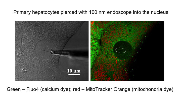
The research led by Dr. Yury Gogotsi, professor of materials science and engineering and director of the A.J. Drexel Nanotechnology Institute (DNI) , and Dr. Gary Friedman, professor of electrical engineering, uses the nanotube-based device, known as a cellular endoscope, to evaluate cells about a thousand times smaller than a human hair. The cellular endoscope interrogates the intracellular environment of living cells, delivers fluorescent quantum dots and analyzes molecules inside a cell without the cell recognizing the needle’s presence.
“Drexel’s W. M. Keck Institute for Attofluidic Probes now manufactures the smallest endoscopes ever created,” Gogotsi said. “Endoscopes provide a potentially transformative technology for studying the fundamentals of single living cells and more broadly, for cell biology.”
Cell biologists usually destroy a large number of cells to extract cellular components and biological molecules needed for identifying diseases and analyzing effects of new drugs, or to achieve a better understanding of how the cell functions. Glass pipettes are widely used to inject material into cells. The pipettes cause too much damage to remain within the cell for a long time and are not designed to report information in the form of optical or electrical signals from within the cell.
“We had an idea for a minimally invasive cellular probe, the tip of which could remain within the cell for a long time while reporting important information in the form of optical and electrical signals and transferring tiny amounts of material to and from the cell. This probe is similar to an endoscope employed by doctors to perform minimally invasive operations inside human patients, only much smaller” said Friedman. “A cellular endoscope reported here is a novel, but conceptually simple device,” said Riju Singhal, a doctoral candidate and author of the article “Multifunctional carbon-nanotube cellular endoscopes” published in the Nature Nanotechnology journal.
“It consists of a single carbon nanotube connected to the tips of larger glass micropipettes that are commonly employed in biological studies, enabling them to become widely used in the near future,” said Singhal.
Dr. Michael Schrlau, research assistant professor in Drexel’s Material Science and Engineering who directs the research laboratory of the W. M. Keck Institute, said, “We’re now building upon the multiple demonstrated functions of cellular endoscopes to help answer elusive cell biological questions. One application of cellular endoscopes being actively pursued is intracellular surface-enhanced Raman spectroscopy with gold-coated endoscopes.”
The Drexel team is funded by the Nanoscale Interdisciplinary Research Team National Science Foundation grant and the W. M. Keck Foundation.
Reference:
Drexel Researchers Create Early Disease Detection and Drug Delivery Device for Single Living Cells// http://www.drexel.edu/news/headlines/drexel-researchers-create-early-disease-detection-and-drug-delivery-device-for-single-living-cells.aspx

 Our collaborative work on porous Ti₃AlC₂ MAX phase for efficient Ti₃C₂Tₓ MXene synthesis has been ranked among the Top 10 most cited papers in the International Journal of Applied Ceramic Technology (IJACT).
 We highly recommend checking out new important paper: “Critical Assessment of Intrinsic Antibacterial Properties and Photothermal Therapy Potential of MXene Nanosheets.” Along with the key findings, we’re also excited to share the Supplementary Cover Art — it beautifully illustrates our vision of MXene-based targeted complexes that can eliminate bacteria via photothermal conversion under near-infrared irradiation.
 Do MXene nanosheets possess intrinsic antibacterial activity? A systematic study of high-quality Ti-, V-, and Nb-based MXenes reveals negligible inherent antimicrobial effects while highlighting their strong potential for targeted photothermal antibacterial therapy.
 Highlights
") We are excited to share that our Carbon-Ukraine (Y-Carbon LLC) company participated in the I2DM Summit and Expo 2025 at Khalifa University in Abu-Dhabi! Huge thanks to Research & Innovation Center for Graphene and 2D Materials (RIC2D) for hosting such a high-level event.It was an incredible opportunity to meet brilliant researchers and innovators working on the next generation of 2D materials. The insights and energy from the summit will definitely drive new ideas in our own development.
We are excited to share that our Carbon-Ukraine (Y-Carbon LLC) company participated in the I2DM Summit and Expo 2025 at Khalifa University in Abu-Dhabi! Huge thanks to Research & Innovation Center for Graphene and 2D Materials (RIC2D) for hosting such a high-level event.It was an incredible opportunity to meet brilliant researchers and innovators working on the next generation of 2D materials. The insights and energy from the summit will definitely drive new ideas in our own development. Carbon-Ukraine team had the unique opportunity to visit XPANCEO - a Dubai-based deep tech startup company that is developing the first smart contact lenses with AR vision and health monitoring features, working on truly cutting-edge developments.
 Our Carbon-Ukraine team (Y-Carbon LLC) are thrilled to start a new RIC2D project MX-Innovation in collaboration with Drexel University Yury Gogotsi and Khalifa University! Amazing lab tours to project collaborators from Khalifa University, great discussions, strong networking, and a wonderful platform for future collaboration.
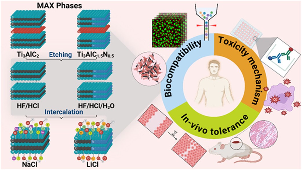 MXenes potential applications include sensors, wound healing materials, and drug delivery systems. A recent study explored how different synthesis methods affect the safety and performance of MXenes. By comparing etching conditions and intercalation strategies, researchers discovered that fine-tuning the surface chemistry of MXenes plays a crucial role in improving biocompatibility. These results provide practical guidelines for developing safer MXenes and bring the field one step closer to real biomedical applications.
. 2D MXenes in the design of heavy metal ion sensors (Review). Trends in Environmental Analytical Chemistry, 47, e00270. DOI:10.1016/j.teac.2025.e00270") An excellent review highlighting how MXene-based sensors can help tackle one of today’s pressing environmental challenges — heavy metal contamination. Excited to see such impactful work moving the field of environmental monitoring and sensor technology forward!
An excellent review highlighting how MXene-based sensors can help tackle one of today’s pressing environmental challenges — heavy metal contamination. Excited to see such impactful work moving the field of environmental monitoring and sensor technology forward!
 Carbon-Ukraine team was truly delighted to take part in the kickoff meeting of the ATHENA Project (Advanced Digital Engineering Methods to Design MXene-based Nanocomposites for Electro-Magnetic Interference Shielding in Space), supported by NATO through the Science for Peace and Security Programme.
 Exellent news, our joint patent application with Drexel University on highly porous MAX phase precursor for MXene synthesis published. Congratulations and thanks to all team involved!
 Our team was very delighted to take part in International Symposium "The MXene Frontier: Transformative Nanomaterials Shaping the Future" – the largest MXene event in Europe this year!
  Last Call! Have you submitted your abstract for IEEE NAP-2025 yet? Join us at the International Symposium on "The MXene Frontier: Transformative Nanomaterials Shaping the Future" – the largest MXene-focused conference in Europe this year! Final Submission Deadline: May 15, 2025. Don’t miss this exclusive opportunity to showcase your research and engage with world leaders in the MXene field!